Renal Cell Carcinoma Liver Metastases Mri

They are the cells from the part of the body where the primary cancer began for example cancerous breast colon or lung.
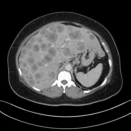
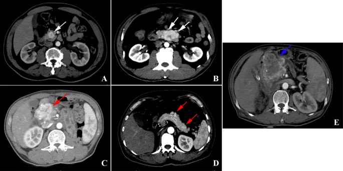
Renal cell carcinoma liver metastases mri. Breast carcinoma hepatic metastases can be hypervascular but these represent a small minority of breast cancer liver metastases. Metastases that are considered hypervascular include renal cell carcinoma carcinoid neuroendocrine tumors thyroid carcinoma and melanoma and have been shown to be better seen on ha phase imaging. Imaging also is a useful tool to see if a particular treatment is working. Homogeneously hypervascular liver metastases from breast are considered rare 3 l.
Ct often the first line of imaging contrast enhanced ct was previously thought to be equivalent to mri for the detection of metastases. Diagnosis may involve ultrasound. Clin cancer res 2008. Metastatic renal cell carcinoma is kidney cancer that has spread to your lymph system bones or other organs.
Mri the appearance of liver metastases on mri is also variable but mri is more sensitive than ct for the detection of liver metastases 5. Liver metastases occur when cancer spreads to the liver from another part of the body. Magnetic resonance imaging measured blood flow change after antiangiogenic therapy with ptk787 zk 222584 correlates with clinical outcome in metastatic renal cell carcinoma. Learn more about liver.
Metastases that hemorrhage includes melanoma renal cell carcinoma choriocarcinoma thyroid cancer lung and breast cancers. Staging tests for kidney cancer may include additional ct scans or other imaging tests your doctor feels are appropriate. Once your doctor identifies a kidney lesion that might be kidney cancer the next step is to determine the extent stage of the cancer. The list of differential diagnoses includes.
Prognosis depends on how far the cancer has spread. There are several tumors which are noted to cause hypervascular metastases. The cancer cells found in a metastatic liver tumor are not liver cells. Follow up imaging after treatment is typically done with ct with dual phase imaging of the abdomen advocated to maximize the detection of solid organ metastases 9.


